Sunday, December 21, 2014

REPOST: Common glaucoma drug reduces risk of vision loss


A new study supports the benefits of widely used glaucoma drug. This article has the details.


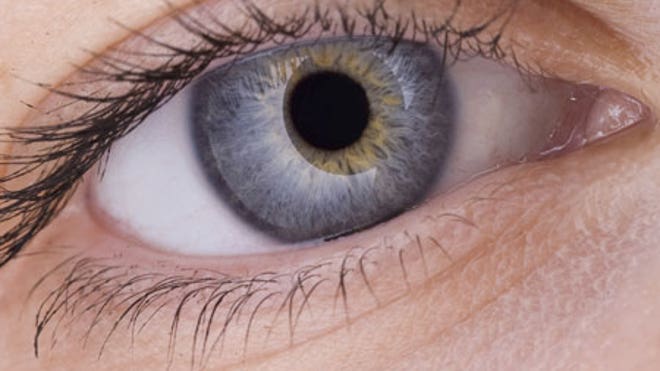
eye-strain
Image Source: thehealthsite.com


A pressure-lowering eye drop has been found to greatly reduce the risk of vision loss in people with open angle glaucoma (OAG), one of the leading causes of blindness.

The trial to assess the vision-preserving effect of prostaglandin analogue eye drop, which is already in common use for treatment of glaucoma, confirmed its efficacy.

"Our findings offer solid proof to patients and practitioners that the visual deterioration caused by glaucoma can be reduced using this treatment," said lead author David Garway-Heath from UCL (University College London) Institute of Ophthalmology.

OAG currently affects around 45 million people worldwide.

Vision loss from glaucoma occurs when the optic nerve is damaged. In most cases, increased pressure inside the eye (intraocular pressure), is thought to contribute to this damage.

"Medication to lower raised eye pressure has been used for decades as the main treatment for OAG to delay progressive vision loss," Garway-Heath explained.

"But, until now, the extent to which the most frequently prescribed class of pressure-lowering drugs (prostaglandin analogues) have a protective effect on vision was not known," Garway-Heath added.

The study recruited 516 newly diagnosed, previously untreated individuals with OAG in Britain.

Half were randomly assigned to daily pressure-lowering eye drops and the other half to a matching placebo.

The risk of visual deterioration was over 50 percent lower in the group treated with daily pressure-lowering eye drops compared to those using placebo drops over 2 years.

The study was published in the journal The Lancet.

Like this Dr. Hitesh Patel Facebook page for more links about revolutionary eye treatments.

Monday, November 24, 2014

REPOST: Games and apps help to improve student's vision

Gaming has always been seen as something that eads to poor eyesight, but the same cannot be said to some games and apps today. This article from Hillsdale Daily News talks about Carson McVey, a first-grader who plays games to improve his eyesight.

With a smile on his face Pittsford Elementary School first-grader Carson McVey plays Bingo with Lisa Pelc, Hillsdale Intermediate School District impaired-orientation and mobility specialist. Image Source: hillsdale.net

With a big smile on his face Pittsford first-grader Carson McVey, 6, yelled "Bingo!" after covering all of his numbers.

With the "Bingo!" McVey received a high-five from Lisa Pelc, Hillsdale Intermediate School District impaired-orientation and mobility specialist. Pelc, who has worked with McVey since preschool, said the game helps McVey with his scanning and number recognition.

The Bingo game is just one of the many exercises the two work through during their weekly sessions to help improve McVey's eyesight.

McVey had cataract surgery last year to improve his vision. Before the surgery he was forced to use a magnifying globe to read and he couldn't see things far away.

"He is getting so good with his reading," Pelc said.

Since the surgery McVey, who now wears bifocals, can read letters from across the room and up close.

Pelc said she is amazed by the changes she has seen in his vision.

Using an iPad outfitted with several apps dedicated to improving vision, Pelc helps McVey work on his reading, sight words, spelling and color recognition.

With a nod McVey said he enjoys using the iPad for learning.

Pelc said all of the new apps for the iPad have been very beneficial to the students she works with on a daily basis. Several of the apps are also aligned with Michigan's Common Core requirements.
Pelc works with approximately 13 students on a weekly basis at several of the schools in the county. She also checks-in on other students who use vision tools in the classroom.

Pelc stressed the importance of catching vision problems at an early age in children to get them corrected. Early detection and treatment can improve a child's learning as they enter school.


Edison-based ophthalmologist Dr. Hitesh K. Patel founded the Patel Eye Associates to treat various  eye conditions. Follow this Facebook page for more information on eye care and treatment.

Friday, October 17, 2014

REPOST: Stem Cells Show Potential Benefits for Eye Diseases


A Massachusetts biotech firm reported that a treatment based on embryonic stem cells might help restore vision. This article from the Wall Street Journal further explains this study.


Image Source: ft.com


Vision Improves for Some Patients in Clinical Trial

Researchers have used stem cells from human embryos to treat patients suffering from severe vision loss, the first time the technique has been shown to be both safe and potentially effective in a sustained way.

The clinical trial, done in 18 U.S. patients, is an important advance in the quest to get the controversial treatment to the clinic—a road marked by years of ethical concerns and legal and political battles.

The latest trial was designed to test whether the stem-cell approach was safe and didn’t lead to tumors or other serious side effects. On that score, the trial was successful.

“It has come through with flying colors,” said Chris Mason, professor of regenerative medicine at University College London, who wasn’t involved with the study.

Given the small number of participants and lack of controls, it is harder to ascertain whether the treatment is broadly effective. Nonetheless, some participants seemed to benefit.

One patient was a 75-year-old rancher from Kansas whose eyesight improved to the point where he could ride a horse again. Another patient went to the mall for the first time. Another traveled to the airport by herself.

“We’ve been hearing about embryonic stem cells for a long time,” said Robert Lanza, chief scientific officer of Advanced Cell Technology Inc., ACTC +3.31% of Marlborough, Mass., and co-author of the study, published Tuesday in the journal Lancet. “We needed some news to show this technology is indeed safe and can help people.”

Embryonic stem cells are master cells that can become all the other cells in the body. The hope is that they can be manipulated to create fresh tissue to transplant into patients and thus treat a range of ailments, from Alzheimer’s to strokes.

It is a controversial approach. Some people oppose removing stem cells from a human embryo because it gets destroyed in the process. They believe that is tantamount to destroying a life.

The stem cells in the trial were used to treat two debilitating eye diseases. One, dry age-related macular degeneration, or AMD, is responsible for more than 80% of the blindness in the developed world. The other, Stargardt’s disease, is the most common form of juvenile blindness and afflicts more than 25,000 people in the U.S. alone.

Both involve the retina—a layer of tissue inside the eye that houses two kinds of light-sensitive cells, rods and cones. There is another layer called the retinal pigment epithelium, or RPE.

The job of RPE cells is to nourish and support the rods and cones. When either of these diseases strikes, the RPE cells stop functioning properly, causing the rods and cones to deteriorate and leading to a progressive loss of vision.

In their trial, Dr. Lanza and his colleagues first obtained an eight-cell embryo from a fertility clinic. (The embryo was left over from fertility treatments and was destined for destruction.) They extracted a single cell, multiplied it in a lab dish and converted some of the resulting cells into RPE cells. The lab-made RPE cells were then injected into the eye.


Transplanted cells appear as a dark spot on the retina of a person with macular degeneration. | Image Source: technologyreview.com


 Half the patients suffered from dry AMD, and half had Stargardt’s. For each patient, the lab-made cells were injected in the eye with the poorer vision. The median follow-up period was 22 months. The trial demonstrated that even after several months, the transplanted cells remained in place and didn’t trigger adverse immune reactions or tumors.

Vision tests suggested that 10 of the 18 treated eyes had improved sight, with eight patients reading more than 15 additional letters on a reading chart in the first year after transplant. Visual acuity remained the same or improved in seven patients, though it decreased by more than ten letters in one patient.

Overall, “we’re seeing a visual improvement,” said Dr. Lanza of Advanced Cell, which funded the study. “That was not what we were expecting—it’s frosting on the cake.”

What could account for the apparent change in vision? Dr. Lanza suggests that some rods and cones in the eye might remain dormant and the lab-made RPEs recharge them so that they function properly.

The research still has a long way to go. Because there were no controls, such as placebo injections in the patients’ eyes that weren’t treated, it isn’t clear whether the improved vision was because of the treatment or some other factor. To help answer that question, the scientists plan to launch a larger trial by the end of the year involving 100 Stargardt’s patients and several dozen who suffer from dry AMD.

Dr. Hitesh K. Patel established Patel Eye Associates to different eye conditions, including myopia, hyperopia, and glaucoma. Visit this Facebook page for more updates.

Monday, September 29, 2014

Eye care through healthy eats: Three nutritious foods for optimal vision

Just like any other part of the body, the eyes need proper nourishment, too. Apart from carrots, there are several other common foods that promote healthy eyesight. The following are some examples:

Kale

Image Source: doitdelicious.com

It's common knowledge that carrots contain beta-carotene, a carotenoid that easily converts into Vitamin A for good vision and overall health improvement. However, Kale and other leafy vegetables such as spinach and collard greens also contain carotenoids, specifically lutein and zeaxanthin. These two carotenoids are the only ones found naturally in the lens and retina of the eye. Studies show that eating food rich in lutein and zeaxanthin helps prevent the onset and slow the progression of cataracts and age-related macular degeneration (AMD) by preventing light-induced damage to the eyes.

A plus: kale also provides vitamins C, K, and B-6; calcium, magnesium, iron, and protein.

Salmon

Image Source: greenretaildecisions.com

Salmon contains large amounts of Omega-3 fatty acids. Studies suggest that the fish helps prevent macular degeneration and dry eye syndrome, as well as decreases the risk of glaucoma and ocular hypertension, a condition that causes abnormal pressure in the eyes which, if left untreated, leads to permanent vision loss in some people.

Those who don't like salmon can try substituting with fish oil supplements.

Almonds

Image Source: healthmag.ir

Almonds are a good source of vitamin E, which has been shown to reduce the risk of advanced AMD by 25 percent for those already exhibiting the beginning symptoms. Other studies suggest that vitamin E prevents or delays the formation of cataracts. It can also be found in vegetable oils, peanut butter, and sweet potatoes.

Eating a diet rich in the aforementioned foods and using preventative measures, such as wearing sunglasses in the daytime and limiting time in front of the computer, can help keep the eyes healthy and injury free.

Dr. Hitesh K. Patel is an Edison-based opthalmologist who specializes in the diagnosis, prevention, and treatment of cataracts. Learn more about eye care by subscribing to this blog.

Thursday, August 28, 2014

REPOST: Eye Care Experts Stress the Importance of Annual Pediatric Vision Screenings


Dr. Norman Saffra, Director of the Division of Ophthalmology at Maimonides Medical Center, stresses the importance of annual pediatric vision screenings in this article on The Jewish Voice.


Dr. Norman Saffra, Director of the Division of Ophthalmology at Maimonides Medical Center, performs an eyesight screening. | Image Source: jewishvoiceny.com


Annual vision screenings are an essential part of preparing your child for Back-to-School. A child suffering from vision problems is at a disadvantage in the classroom, in sports, at home studying, and more.

“A child’s vision is constantly developing,” notes Dr. Norman Saffra, Director of the Division of Ophthalmology at Maimonides Medical Center. “It is even more important for children currently wearing glasses to be re-evaluated annually as their prescriptions may change as they grow.”

Parents should rely on their pediatricians for initial vision screenings. These screenings will detect common vision problems such as nearsightedness, farsightedness, blurred vision, and other eye abnormalities. If a problem is detected, the next step is to make an appointment with a pediatric eye care specialist for further evaluation and treatment.

“If parents notice a vision issue in between annual check-ups, they should be sure to schedule an appointment as soon as possible,” says Dr. Saffra. “When in doubt, check it out.”

Throughout the school year, take note of symptoms of developing vision problems. Signs may include: difficulty seeing the blackboard or reading books, excessive eye rubbing and increasing headaches. Take special note of children squinting or blinking frequently, or closing one eye when reading. If one eye seems droopy or drifting, your child may be developing a “lazy eye.”

“Not all vision problems are lifelong reasons to wear glasses,” points out Dr. Roman Polishchuk, Pediatric Optometrist at Maimonides Medical Center. “Sometimes as children develop, their eyesight needs minor corrective assistance, after which, your child can stop wearing glasses.”

The Maimonides ophthalmology team strongly urges parents not to miss their child’s annual vision screening.

The Maimonides Infants & Children’s Hospital is Brooklyn’s first and only fully-accredited children’s hospital, offering the full array of pediatric sub-specialties. The inpatient units feature an aquatic theme with fish tanks maintained through a partnership with the New York Aquarium. For more information on the outstanding clinical services provided at the Maimonides Infants & Children’s Hospital, visit our website at http://www.maimonidesmed.org/Pediatrics/MICH.aspx

Edison-based ophthalmologist, Dr. Hitesh K. Patel established the Patel Eye Associates for people in need of eye treatments. Subscribe to this Facebook page to learn more about eye care.

Friday, July 18, 2014

REPOST: Novartis-Google team on lens for diabetes, vision issues

 Among the new technologies in eye care that is in development is the "smart contact lens". Read this article from USAtoday.com for more details.

Image Source: usatoday.com

Google is teaming up with pharmaceutical giant Novartis to develop a "smart" contact lens intended to replace reading glasses for people who can't read without them and glucose monitors for those with diabetes.

For people with vision problems, the device would work like the autofocus of a camera, allowing them to focus on close-up things like the words in a book. It will be designed to work as a contact lens that is changed out regularly, or as an intra-ocular lens, permanently inserted into the eye during cataract surgery.

For diabetics, the lenses would replace regular finger sticks designed to read out a person's blood glucose level. Instead, the lens will "read" glucose levels in tears, sending information wirelessly to a handheld device that will warn patients when they need to eat or lower their glucose levels.

"These are issues that have been unmet medical needs for quite some time," said Novartis CEO Joseph Jimenez.

Novartis' eye-care division Alcon, based in Texas, inked the deal today to do the development work, along with Google X, an innovation lab at the information company's Mountain View, Calif., headquarters, which had previously announced plans to make "smart" contacts.

Jimenez, said he hopes the contact will be the first of many technologies the two companies develop together.

"It was very clear that there could be a very nice synergistic value between bringing high tech together with biology to solve some of the biggest health care issues that we're facing," Jimenez said.

The work is still preliminary, he said, with testing in people expected to begin next year. It will be a few years before the lens can be considered for regulatory approval and reach customers.

He said a price had not been decided for the lenses, and would not disclose how much the two companies will be investing in the project other than to say that it will be "commensurate with the business opportunity."

More than 1.7 billion people worldwide have presbyopia, the medical term for eyes that lose their ability to focus up-close with age, and more than 380 million have diabetes, he said.

Diabetes experts said they see two potential problems with glucose-monitoring lenses.

First, it's not clear whether glucose in tears, which the device would measure, changes in sync with the glucose in blood.

"There are no available data to show any reliability of this sensor," said Robert Ratner, chief scientific and medical Officer of the American Diabetes Association. "Is this going to be a situation where you have to change the contact once a day, once a week or once an hour? All of these are unknowns."

Second, diabetics are most at risk for crashing blood sugar levels at night while they're sleeping. But that's also when most people take out their contacts.

"Any type of contact device is going to need to be something that can be worn overnight if it's going to be able to realize the benefits people need, which is to get warning alarms when their glucoses are going out of range overnight," said Howard Wolpert, director of the Joslin Institute for Technology Translation at the Harvard-affiliated Joslin Diabetes Center in Boston.

For aging eyes, there are also many other alternatives to the contacts, from glasses to low-tech contacts to LASIK surgery, said Kerry Assil, an ophthalmic surgeon in Los Angeles.

But still, he said, if the "smart" contacts can be made to work, it would be a boon to patients. "Any new thing that can provide people with a slightly enhanced visual clarity, reduced side effects and so forth, tremendously elevates their sense of well being and their sense of confidence."

Dr. Hitesh K. Patel is an Edison-based opthalmologist and founder of the Patel Eye Associates. Follow this Twitter account to learn more about the latest trends in eye care.

Monday, June 30, 2014

Age–related macular degeneration: Family history is a factor

Image Source: optegra.com

 It is important to know your family history, particularly when it comes to eye diseases such as age-related macular degeneration (AMD). Studies have suggested that family history is a significant determinant in the development of eye diseases particularly in AMD. The risk increases if the person has an immediate family member, for instance, a parent or a sibling, who already has the disease.

Image Source: hollows.org.au

Macular degeneration is one of the leading causes of vision loss among those aged 50 and above. As the name suggests, it is the degeneration or loss of the macula, which is a small spot near the center of the retina. The macula is the part of the eye needed for sharp, central vision. This allows people to see objects that are before them. Damage to the macula would blur the image. Over time, the blurred image may get larger and the person could develop blank areas in their central vision. Objects also do not appear as bright as they used to be.

 The development of AMD varies from person to person. Some people may not suffer from vision loss until after a long period of time. AMD does not lead to complete blindness. Nevertheless, the loss of central vision in AMD will affect how the person goes through everyday activities, such as driving, reading, writing, and cooking.


Image Source: aoa.org

 Unfortunately, early and intermediate stages of AMD are often without symptoms and go undetected. Only by using a comprehensive dilated exam can a person determine whether one has AMD. Still, AMD may be prevented through the use of proper lifestyle changes. It is also highly recommended to take a thorough audit of one’s family history to surmise potential risks.

Maintaining good eye health and treating eye-related diseases is the mission of ophthalmologist Dr. Hitesh K. Patel of Edison, New Jersey, and the primary reason behind the establishment of Patel Eye Associates. Receive up-to-date information about the latest in eye health by liking this Facebook page.

Saturday, May 31, 2014

New studies asses patient’s adherence to glaucoma medication

A new study reveals that a fairly large number of patients suffering from glaucoma did not regularly use their prescribed eye drops.

Image source: www.atlanticeyeinstitute.com

The research, which was conducted at university-based glaucoma clinic, recruited 407 patients who were undergoing treatment that required daily prostaglandin eye drops. Aimed to measure their medical adherence, the patients were given a container with an electronic cap that recorded each time it was opened.

It was revealed later on that 82.8 percent of the participants took their medication properly on at least 75 percent of days, while the remaining 17.2 percent were not able to adhere to their prescribed medication.

Image source: www.oregoneyecenter.com


Furthering the study, the researchers made their second intervention by including text or voice messages to assist the patients with their eye drop medication.

As the experiment concluded, the researchers found that the adherence rate among those who failed to take their medication in the first batch increased from 53 percent to 64 percent.

Although the barriers to patients’ medication adherence were considered complex, the researchers concluded that the inclusion of text or voice messages can help patients adhere to their eye drop medication.


Image source: www.glaucoma.org


Given these results, the researchers noted that telecommunication-based reminders can help patients and doctors track one’s daily glaucoma medications. They further stressed that this latest discovery is essential because it can potentially support today’s ophthalmology practice.

Dr. Hitesh K. Patel is an Edison, New Jersey-based ophthalmologist. Read more about glaucoma and other vision problems at www.pateleyeassociates.com.

Sunday, March 9, 2014

REPOST: Off with your glasses: Visual crowding linked to retina, brain processing

Tel Aviv University researchers discovered a link between sharp vision and brain's processing speed. Find out how the brain integrate visual formation from this ScienceDaily.com article.
 
 ****

Middle-aged adults who suddenly need reading glasses, patients with traumatic brain injuries, and people with visual disorders such as "lazy eye" may have one thing in common -- "visual crowding," an inability to recognize individual items surrounded by multiple objects. Visual crowding makes it impossible to read, as single letters within words are rendered illegible. And basic cognitive functions such as facial recognition can also be significantly hampered. Scientists and clinicians currently attribute crowding to a disorder in peripheral vision.


Image Source: en.wikipedia.org

Now Prof. Uri Polat, Maria Lev, and Dr. Oren Yehezkel of Tel Aviv University's Goldschleger Eye Research Institute at the Sackler Faculty of Medicine have discovered new evidence that correlates crowding in the fovea -- a small part of the retina responsible for sharp vision -- and the brain's processing speed. These findings, published in Nature's Scientific Reports, could greatly alter earlier models of visual crowding, which emphasized peripheral impairment exclusively. And for many adults lost without their reading glasses, this could improve their vision significantly.

"Current theories strongly stress that visual crowding does not exist in the fovea, that it's a phenomenon that exists only in peripheral visual fields," said Prof. Polat. "But our study points to another part of the eye altogether -- the fovea -- and contributes to a unified model for how the brain integrates visual information."

 A trained eye

According to Prof. Polat, vision is dynamic and changes rapidly, but it takes time for the brain to process this visual information. Rapidly moving tickers on TV, or traffic signs seen as the driver speeds past, are difficult for anyone to read. However, given enough time, someone with excellent vision can fully recognize the words. Those with slower processing speeds -- usually the result of poor perceptive development or age -- may not be able to decipher the tickers or the traffic signs. In the study, Prof. Polat employed his expertise in improving vision by retraining the brain and the foveal part of the eye, using exercises in which speed is a key element.

 
Image Source: flickr.com

"Training adults to reduce foveal crowding leads to improved vision. A similar training we conducted two years ago allowed adults to eliminate their use of reading glasses altogether, using a technology provided by the GlassesOff company. Other patients who had lost sharp vision for whatever reason were also able to benefit from the same training and improve their processing speed and visual capabilities," said Prof. Polat. Maria Lev, who performed the study as a part of her doctoral thesis, said one young subject had experienced significant limitations in school for years and had been unable to obtain a driver's license due to severe visual impairment from foveal crowding. After undergoing training that emphasized a foveal rather than a peripheral focus, he was able to overcome the handicap.

 
Image Source: zmescience.com

 "He finally managed to learn to read properly and found his way forward," said Lev. "I'm proud to say that today he is not only eligible for a driver's license, he's also been able to earn his master's degree."  

Follow this Dr. Hitesh K. Patel Twitter page to get more updates on vision care.

Thursday, February 20, 2014

REPOST: New eye layer has possible link to glaucoma, study shows

How will the new layer that's discovered in the human eye affect eye diseases like glaucoma? Read the answer from this ScienceDaily.com article.
***
A new layer in the human cornea -- discovered by researchers at The University of Nottingham last year -- plays a vital role in the structure of the tissue that controls the flow of fluid from the eye, research has shown.

Image Source: foxnews.com

The findings, published in a paper in the British Journal of Ophthalmology, could shed new light on glaucoma, a devastating disease caused by defective drainage of fluid from the eye and the world's second leading cause of blindness.
The latest research shows that the new layer, dubbed Dua's Layer after the academic Professor Harminder Dua who discovered it, makes an important contribution to the sieve-like meshwork, the trabecular meshwork (TM), in the periphery of the cornea.
The TM is a wedge-shaped band of tissue that extends along the circumference of the angle of the anterior chamber of the eye. It is made of beams of collagen wrapped in a basement membrane to which trabecular cells and endothelial cells attach. The beams branch out randomly to form a 'meshwork'.
Pressure within the eye is maintained by the balance of aqueous fluid production by eye tissue called the ciliary body and drainage principally through the TM to the canal of Schlemm, a circular channel in the angle of the eye.
Defective drainage through the TM is an important cause of glaucoma, a condition that leads to raised pressure in the eye that can permanently affect sight. Around 1 to 2% of the world's population yearly have chronic glaucoma and globally around 45 million people have open angle glaucoma which can permanently damage the optic nerve -- 10% of whom are blind.
The latest research by Professor Dua and colleagues in Academic Ophthalmology at The University of Nottingham sheds new light on the basic anatomy of Dua's Layer, which is just 15 microns thick but incredibly tough. Comprised of thin plates of collagen, it sits at the back of the cornea between the corneal stroma and Descemet's membrane.
By examining human donor eyes using electron microscopy, the researchers were able to look at Dua's Layer beyond the central part of the cornea to shed more light on its features at the extreme periphery of the cornea. They discovered that the collagen fibres of Dua's Layer also branch out to form a meshwork and that the core of TM is in fact an extension of Dua's Layer.
It is hoped the discovery will offer new clues on why the drainage system malfunctions in the eyes of some people, leading to high pressure.
Professor Dua said: "Many surgeons who perform lamellar corneal transplant recognize this layer as an important part of the surgical anatomy of the cornea. This new finding resulting from a study of the microanatomy of the periphery of the layer could have significance beyond corneal surgery."
The breakthrough discovery of Dua's Layer was first revealed in a paper in the academic journal Ophthalmology in June last year and was widely covered by the world's scientific and lay media. The paper became the number one downloaded ophthalmology paper from the website ScienceDirect between July and September 2013 and was ranked the 11th most downloaded from the website for the whole of medicine and dentistry.

***
Dr. Hitesh K. Patel is the founder of the Edison-based eye center, Patel Eye Associates. Know more about him by following this Twitter account.

Thursday, January 16, 2014

REPOST: Cause of destructive cascade of diabetic retinopathy

This MedicalNewsToday.com article discusses the roles  and effects of excessive ROS or reactive oxygen species in our body and how will it affect our vision.


The retina can be bombarded by reactive oxygen species in diabetes, prompting events that destroy healthy blood vessels, form leaky new ones and ruin vision.

Now researchers have learned that those chemically reactive molecules must come from both the bone marrow as well as the retinal cells themselves to cause such serious consequences.

"It's a cascade that requires two players to signal the next event that causes the damage," said Dr. Ruth Caldwell, cell biologist at the Vascular Biology Center at the Medical College of Georgia at Georgia Regents University.

The good news is the finding also provides two new points for intervention, said Dr. Modesto Rojas, MCG postdoctoral fellow and first author of the study in the journal PLOS ONE.

Excessive glucose in the blood prompts excessive production of reactive oxygen species, or ROS, and the light-sensitive retina is particularly vulnerable. Caldwell's research team had previously documented that ROS from white blood cells produced by the bone marrow as well as from retinal cells were the major instigators in diabetic retinopathy, a leading cause of blindness worldwide. But they weren't sure which mattered most.

So they looked as several different scenarios, including mice lacking the ability to produce ROS by either the retinal or white blood cells, and found that if either were lacking, future damage was essentially eliminated. "One alone can't do it," said Caldwell, the study's corresponding author. "They did not develop the early signs of diabetic retinopathy that we were measuring."

While blocking ROS production by retinal cells could be difficult, drugs already exist that reduce activation of white blood cells. Those cells not only make ROS, but also adhere to blood vessel walls in the retina that become sticky in diabetes, Rojas said. In fact, a study published in October 2013 in PLOS ONEshowed that neutrophil inhibitory factor could block the vascular lesions that are a hallmark of diabetic retinopathy without hurting the immunity of diabetic mice. The MCG scientists note that decreased activation does not impact the immune protection white blood cells also provide.

Next steps include studying those drugs in their animal models and learning more about how ROS causes the collateral damage that can destroy vision. "All of this is some sort of wound-healing response gone wrong," Caldwell said.

ROS, a natural byproduct of the body's use of oxygen, has healthy roles in the body, including cell signaling, but is destructive at high levels that result from disease states such as diabetes.


Dr. Hitesh K. Patel, an ophthalmologist, serves Edison, New Brunswick and all areas of Middlesex County, NJ. LASIK surgery is one of his specializations.Get more updates by following this Twitter page.